3D Printed Parotid Gland and Facial Nerve Dissection
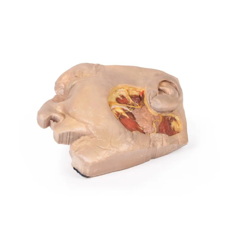
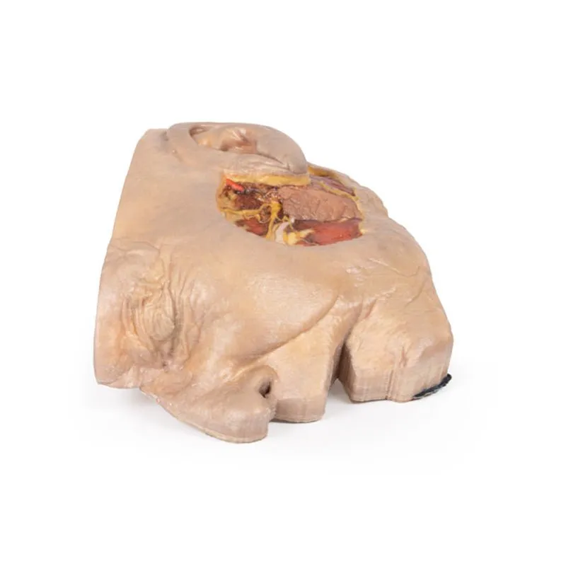
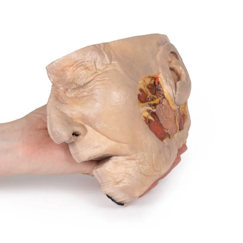
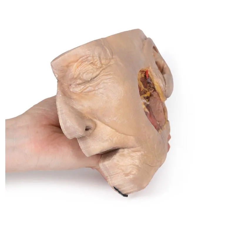
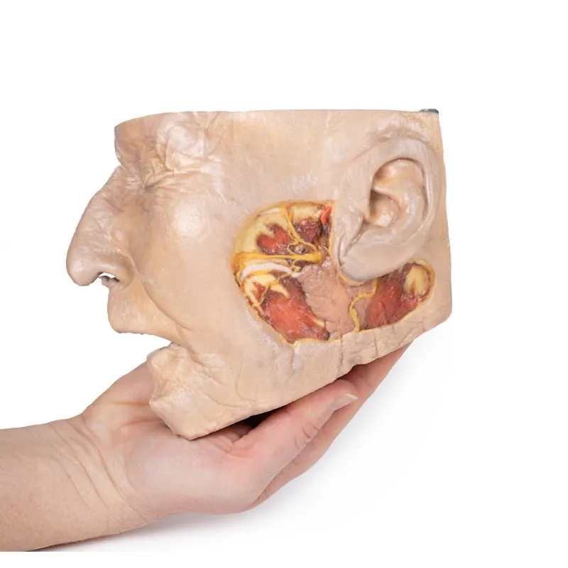
This 3D model provides a superficial dissection window into the lateral face to demonstrate the anatomy of the
parotid gland relative to surface features and neurovascular structures. These structures are of particular
significance for management in Mohs surgery in the management of skin cancers, or in certain plastic and
reconstructive surgical procedures.
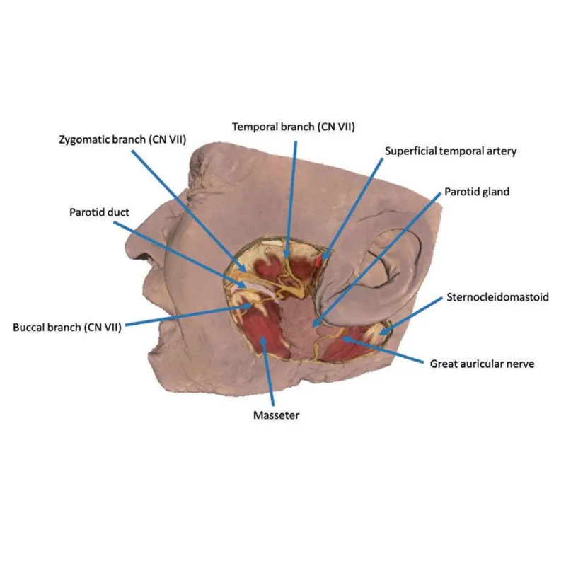
The opened window extends from just anterior to the external ear, from the
level of the zygomatic arch to the angle of the mandible and extending from the anterior margin of the masseter
muscle to the origin of the sternocleidomastoid muscle. Exposed within the window is the bulk of the parotid gland,
with the superior portions of the gland removed to demonstrate the superficial temporal artery and the facial nerve
dividing into the superior terminal branches (e.g., the temporal, zygomatic and buccal). The parotid duct traverses
the opened dissection window before passing towards the buccal region (and its termination into the buccinator
muscle). An ascending branch of the great auricular nerve can be observed along the inferior and posterior margins
of the parotid gland, and just anterior relative to the sternocleidomastoid.
GTSimulators by Global Technologies
Erler Zimmer Authorized Dealer


